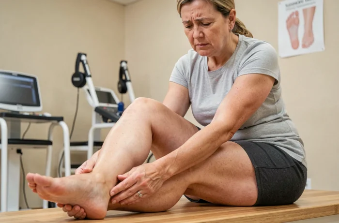
A runner resumes training after an ankle sprain and, a few weeks later, experiences persistent burning along the outer edge of the foot. The calf seems normal and mobility is good, but the area between the outer ankle bone and the little toe remains painful at the slightest shoe friction. This pattern often points to sural nerve involvement, affecting a purely sensory nerve whose irritation frequently goes unnoticed.
Shoes and scars: two mechanical pitfalls for the sural nerve
The sural nerve runs along the back and outer side of the calf, passes behind the outer ankle bone and ends along the lateral edge of the foot, extending to the fifth toe. Its superficial course makes it vulnerable to repeated local pressure.
The role of footwear is often underestimated. A stiff upper, a high-top boot pressing against the outer ankle bone, or an overly tight heel counter can compress the nerve over several centimetres. Professional safety footwear and some hiking boots are particularly common culprits.
Another frequent pitfall is scar tissue compressing the nerve after ankle surgery. Surgery on the Achilles tendon, fixation of a calcaneal fracture, or repair of the peroneal tendons can lead to fibrous tissue forming around the nerve. The risk varies depending on the surgical technique: some percutaneous Achilles tendon repairs increase the likelihood of nerve symptoms compared with open procedures, because the operation is performed blindly in an area where the nerve passes very close by.
When investigating sural nerve pain in the foot, recurrent ankle sprains should also be considered. Stretching or a direct impact during the initial injury can damage the nerve, while residual instability can then keep irritating it with every misstep.

Sural nerve symptoms: distinguishing burning, numbness and mechanical pain
The sural nerve has no motor function. It does not control any muscles in the foot or leg. An isolated injury therefore causes neither loss of strength nor a limp due to paralysis. This is a useful clue when assessing the cause: if the foot is “dropping” or the toes no longer respond, the problem is likely elsewhere.
The characteristic signs affect sensation:
- Burning or electric-shock sensations along the outer edge of the foot, sometimes extending up towards the calf
- Numbness or a “cardboard-like” feeling in the skin between the outer ankle bone and the little toe
- Pain that worsens when wearing closed shoes, walking for a long time, or applying direct pressure to the area behind the outer ankle bone
- Tingling that occurs at rest, especially at night, when the position of the foot keeps the nerve compressed
The precise location helps distinguish sural nerve pain from other causes of pain on the outer side of the foot. Peroneal tendinitis causes deeper pain that worsens with active foot eversion. A stress fracture of the fifth metatarsal causes a distinct, localised point of bone tenderness. The sural nerve causes superficial, burning pain, often triggered by something as simple as contact with the sock fabric.
Diagnosing sural neuropathy: what tests reveal—and what they miss
A clinical examination is the first step. The clinician taps or presses along the nerve behind the outer ankle bone to reproduce the symptoms (Tinel’s sign). Comparing sensation in both feet can help identify a sensory deficit.
A normal nerve conduction study does not rule out localised irritation. Electromyography measures the nerve’s overall conduction speed, but intermittent or highly localised compression may not affect the signal enough to be detected. Results should always be interpreted alongside the symptoms reported and the physical examination.
High-resolution ultrasound is a valuable addition when assessing superficial nerve injuries. It can reveal nerve thickening, scar tissue surrounding the nerve, or a compressive mass. MRI is used when there is concern about surrounding tissues or a deeper injury that ultrasound cannot assess.
What imaging cannot replace
No imaging test can reproduce the patient’s pain. Ultrasound abnormalities are regularly found in people without symptoms, while some patients with significant discomfort have nerves that look normal. Sural neuropathy is primarily a clinical diagnosis; additional tests are used to confirm it, pinpoint its location, or rule out a specific structural cause.
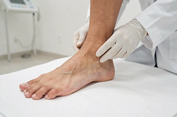
Relieving sural nerve pain in the foot: gentle mobilisation and footwear adjustments
The first step, and often the most effective, is to remove the source of mechanical compression. Changing shoes, choosing a style with a soft upper and no rigid heel counter pressing against the outer ankle bone, or simply adding protective padding over the area that rubs can reduce pressure on the nerve.
Nerve-gliding exercises are used in rehabilitation to help restore the nerve’s ability to move through the surrounding tissues. They involve gentle, progressive movements of the ankle and foot combined with knee flexion. Evidence specific to the sural nerve remains limited, and overly forceful mobilisation can worsen burning and numbness. It is best to practise under supervision, at least during the first sessions.
Medical treatments vary depending on the severity:
- Anti-inflammatory medication and neuropathic pain relievers to reduce inflammation and electric-shock sensations
- A local corticosteroid injection when the compression has been clearly identified and conservative treatment is not enough
- Surgical neurolysis as a last resort, to free the nerve from scar tissue or other compressive tissue, with careful identification of the nerve’s course
The sural nerve regenerates slowly after direct injury. Axonal regrowth progresses by only about one millimetre per day, meaning that sensory recovery after a nerve transection or decompression surgery takes months. Patience is as much a part of treatment as the exercises.
Monitoring symptoms during the first few weeks after changing footwear or starting rehabilitation provides a reliable indication of progress. If the burning gradually subsides, conservative treatment is working. If it worsens or the numbness spreads, further assessment with ultrasound or electromyography can help reassess the situation before prolonged compression compromises nerve recovery.